Bridging the Gap in Anatomical Education for Implantologists
Anatomical textbooks and atlases often fall short of meeting the clinical needs of oral implantologists by focusing excessively on intricate details that can overwhelm practitioners.
Certain anatomical landmarks are challenging to depict in diagram form, leading to confusion for students and professionals when they encounter actual specimens in the dissection lab or operating room.
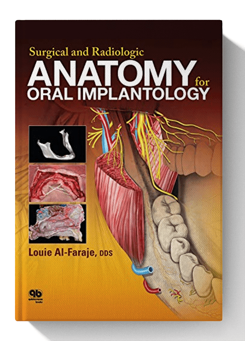
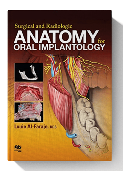
This book addresses these challenges by illustrating the structures of the maxilla, mandible, and nasal cavity as they exist in real cadaver specimens and clinical cases.
Several chapters feature full-page images of specific cadaver sections, with all relevant anatomical parts clearly labeled for easy reference.
It also includes cone beam computed tomography (CBCT) images, demonstrating how this technology can assess bone density, the width of the alveolar ridge, and the precise distances available for implant placement in relation to anatomical landmarks prior to selection.
Overall, this book aims to simplify the learning and execution of implant-related surgical procedures in regions of the body that present unique topographic and anatomical challenges.


Reviews
There are no reviews yet